Anchor ID #services

Radiology Services

Using state-of-the-art technology designed especially for infants, children and teens, Connecticut Children’s radiologists can obtain accurate, extremely detailed images while your child remains safe and comfortable. We adhere to the national Image Gently standards to limit radiation exposure to children while still providing safe, high quality pediatric imaging. Our team of radiologists and technologists provide comprehensive radiology and imaging services to help evaluate an array of pediatric conditions.

Accredited by the American College of Radiology for computed tomography, MRI and ultrasound, Connecticut Children’s exceeds national standards for quality assurance and safety guidelines.

Our state-of-the-art imaging equipment is appropriate for necessary tests and treatments and our radiologists are well qualified to perform and interpret medical images and administer therapy treatments that use radiation.

Our radiologists use an array of imaging procedures to create a picture of the inside of a pediatric patient’s body, including some that use medical radiation and others that do not. MRI and ultrasound do not use medical radiation and may or may not provide the kind of information needed to assess and diagnose your child’s condition. Other tests that do use radiation, such as a CT scan, can be used to provide a better image of internal conditions to the physician, and are always carried out at the lowest possible dosage, for infants, children and teens.

Bone Density is measured using DEXA (dual-energy X-ray absorptiometry) technology, which evaluates bone mineral density using two low-dose X-ray beams aimed at patients’ bones. DEXA scans are painless and are typically used to determine and track bone strength of the spine, hip and wrist.

Computed Tomography (CT) uses X-ray beams to produce 3D, cross-sectional images of specific areas of the body including the head, abdomen and extremities.

The test might require contrast—a special liquid dye that helps radiologists see the patient’s body better—and/or sedation in order to keep the child completely still. Depending on the part of the body being scanned, the contrast may be given by mouth or through a needle placed in a vein in the hand, arm or foot.

Patients lay very still on a narrow bed that moves slowly into a tube, which scans the body painlessly.

Fluoroscopy uses a continuous X-ray beam to obtain a sequence of images of the structures inside the body which are projected on a monitor to show real-time organ motion.

Before fluoroscopy scans, patients are given a liquid contrast material, which helps make the images clearer. This technique is helpful for visualizing parts of the gastrointestinal and urinary tracts, including the esophagus, stomach, small and large intestine, rectum, kidneys, ureter, bladder and urethra.

Connecticut Children’s offers VCUG, barium enema, UGI, esophagram, PICC insertion and other types of fluoroscopy.

Interventional radiology uses images to help guide minimally invasive procedures for diagnostic or treatment purposes. The images enable interventional radiologists to locate disease and navigate to it using tiny instruments.

Magnetic Resonance Imaging (MRI) uses a very strong magnet and radio waves to take a series of detailed images that provide multidimensional views of soft tissues of the extremities, body, brain and spine.

MRI is a painless, noninvasive test that does not use radiation.

Ultrasounds use sound waves to form images of the inside of the body. The ultrasound transducer is placed directly onto the skin, and sends sound waves that bounce off organs to make an image on the screen.

Ultrasound is best used when more information is needed about soft tissue organs such as the kidney, bladder, liver, spleen, pancreas, gallbladder, uterus, and ovaries. This test may also be used on infants to check for dislocated hip joints or to see the brain of a newborn.

X-ray (radiography) sends low levels of radiation through the tissues and bones to provide images of the inside of the body.

After every imaging procedure, a pediatric radiologist will review the images and then discuss the results with patients and families, who may then request a copy of the image.

Images are available electronically to Connecticut Children’s referring providers and upon request to community pediatricians and other primary care physicians. Reports are sent to all referring providers.

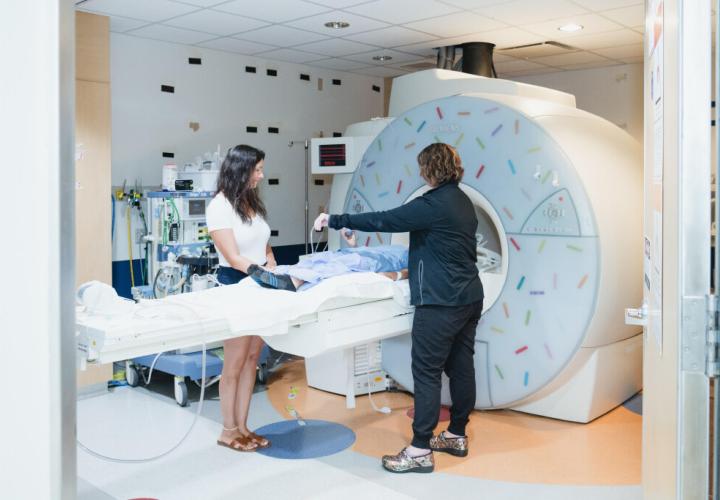
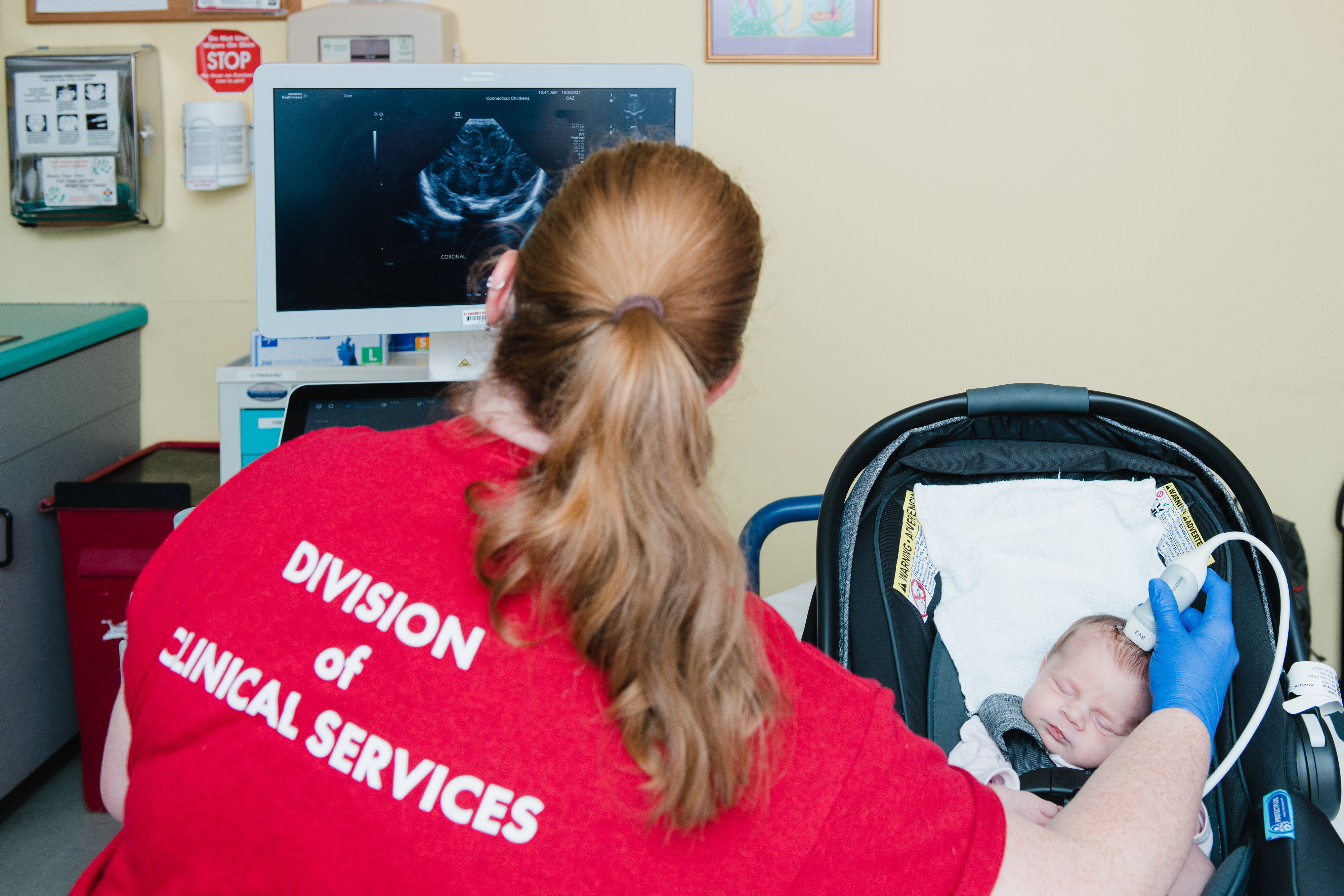
Specializing in Pediatric Radiology

Our highly skilled, board-certified pediatric radiologists are dedicated to the diagnosis and treatment of childhood illnesses using equipment such as X-rays, magnetic resonance imaging (MRI) and computed tomography (CT) to make images of inside the body.

Learn more about our team
Anchor ID #locations

Pediatric Radiology Locations

To refer a child for imaging services at Connecticut Children’s Medical Center, please complete the Clinical Services Referral Form.

Exterior view of Connecticut Children's Medical Center in Hartford

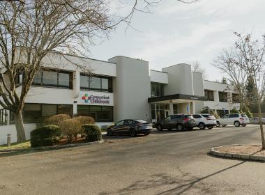
Connecticut Children’s Medical Center – Hartford

282 Washington Street
Hartford, CT06106
United States

Exterior view of 310 Western Boulevard in Glastonbury

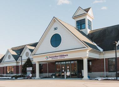
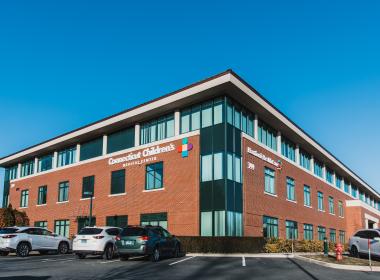
Connecticut Children’s Specialty Care Center – Glastonbury

310 Western Boulevard
Glastonbury, CT06033
United States

Exterior view of 105 Newtown Road in Danbury

Connecticut Children’s Specialty Care Center – Danbury

105-A Newtown Road
Danbury, CT06810
United States

Connecticut Children’s Specialty Care Center – Westport

191 Post Road West
Westport, CT06880
United States

Exterior view of 399 Farmington Ave in Farmington

Connecticut Children’s Specialty Care Center – Farmington (399 Farmington Ave.)

399 Farmington Avenue
Farmington, CT06032
United States

Anchor ID #resources