What is a fetal echocardiogram?
A fetal echocardiogram, also known as a fetal echo, uses sound waves to make images of an unborn baby’s heart.
This safe, simple procedure reveals really important information: Experts can see not only the structure of the heart, but how well it’s functioning. They can often diagnose congenital heart defects long before a baby is born.
Why would you need a fetal echo?
During pregnancy, you could be referred for a fetal echo for a number of reasons.
For example, you might need a fetal echo if you have or have had:
- A family history of congenital heart defects
- In vitro fertilization (IVF) or other assisted reproductive technology
- Certain medical conditions, such as diabetes, lupus or exposure to certain substancesY
You might also be referred if an OB screening or another test showed a possible problem in the baby, for example:
- Possible problem in the heart
- Possible problem with another major organ system
- Possible genetic or chromosomal abnormality
What happens during a fetal echo?
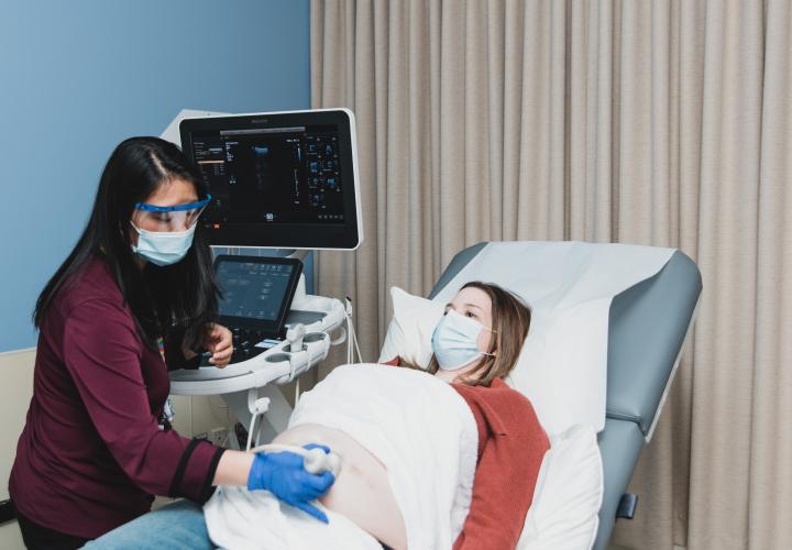
A fetal echo is similar to the routine ultrasounds that you have during pregnancy, but it produces a much more detailed result.
- The procedure is painless and very safe for both mom and baby. It’s performed in a darkened room, while you’re lying down.
- A specialist (known as a fetal sonographer) puts gel on your belly, then moves a wand over it that sends sound waves to the baby’s heart and back again.
- The sound waves are used to create detailed pictures of the baby’s heart.
A fetal echo can take anywhere from 30 minutes to 2 hours, depending on the position that the baby is in. Usually, results are ready the same day.
What else should you expect at your fetal cardiology visit at Connecticut Children’s?
In addition the fetal echo, you’ll receive additional support from our team.
- Care from an expert in fetal cardiology: We take time to explain everything we see in detail and answer all of your questions.
- Introductions to your baby’s future care teams, if needed: This may include specialists from Connecticut Children’s neonatal intensive care unit (NICU), cardiac interventionalists, or pediatric heart surgeons.
- Close communication with your other care teams: We partner closely with other care providers for both baby and mother, like maternal fetal medicine experts, labor and delivery teams and general pediatricians.
- Help coordinating other resources: We can connect you with all the services you need to navigate your baby’s care, from NICU or PICU tours to ongoing emotional support.