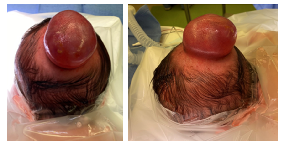
What is a Cephalocele?

A cephalocele is a rare condition where part of the brain or its covering pushes through an opening in the skull. These openings may be present from birth (congenital) or develop later in life (acquired).

There are two main types of cephaloceles:

  1. Meningoceles: These occur when the protective layers around the brain and the fluid that surrounds it, known as cerebrospinal fluid (CSF), push through an opening in the skull.
  2. Encephaloceles: These occur when the protective layers, CSF, and part of the brain tissue push through an opening in the skull.

Encephaloceles are classified based on where they occur on the skull. The two main locations are the posterior (back) and anterior (front) regions. Posterior encephaloceles can be further be classified into:

•    Occipital: at the back of the head
•    Occipitocervical: where the skull meets the neck
•    Parietal: on the top side of the head


Our Care Team

At Connecticut Children’s, we understand how challenging these conditions can be for families. That’s why we have a dedicated multidisciplinary team to care for each patient. Our team is composed of specialists in Neurosurgery, Fetal Surgery, Maternal Fetal Medicine, Neonatology, Pediatric Surgery, and Fetal Radiology, who work together to create a personalized treatment plan. Our goal is to ensure that every patient receives the best possible care and support at every stage of their journey.

For Medical Professionals

Cephaloceles are congenital or acquired malformations that are characterized by herniation of the intracranial contents through a skull defect. Lesions that only contain meninges and cerebrospinal fluid (CSF) are referred to as meningoceles, whereas encephaloceles also contain brain parenchyma (1). Cephaloceles account for 10-20% of all craniospinal dysraphisms and have an estimated prevalence of 0.8-5 per 10,000 live births (2-6). The true incidence is suspected to be higher, as many pregnancies are terminated when the diagnosis is made in utero. The embryology underlying congenital cephaloceles has been debated; some attribute the malformation to disruption of the closure of the neural tube due to the failed separation of the neuroectoderm and surface ectoderm during the 4th week of gestation. More recent evidence has supported the classification of cephaloceles as a post-neurulation disorder, which leads to herniation of the closed neural tube (2, 3, 7).

Encephaloceles are typically classified by location, and include posterior and anterior encephaloceles. Posterior encephaloceles are classified as occipital, occipitocervical, or parietal. Approximately 70-90% of all encephaloceles involve the occipital region, particularly in North America and Europe. Occipital encephaloceles are further characterized by their relationship to the torcula – supratorcular or infratorcular. There is a female predominance, and 90% affect the midline (8). Anterior encephaloceles are more commonly found in Southeast Asia (9). Anterior encephaloceles result from abnormal development of the foramen cecum, and are classified as basal encephaloceles (which herniate through the cribriform plate, ethmoid, or sphenoid sinuses) and sincipital encephaloceles (which include nasofrontal, nasoethmoidal, and nasoorbital subtypes) (2, 10-12). 

Encephaloceles comprise 5% of all severe structural defects of the central nervous system that are detected in utero, and approximately 80% are detected during the first trimester (13-15). Maternal serum alpha-fetoprotein levels are of unclear significance, as encephaloceles are typically skin-covered. Therefore, prenatal imaging is the mainstay of the diagnostic workup.


Sonographic Imaging

Prenatal ultrasound has been used to detect fetal encephaloceles since the 1970s (16). Ultrasound can be used to characterize the size of the cephalocele sac, the composition of the herniated contents (to differentiate meningoceles from encephaloceles), and the size of the underlying bony defect (17, 18). Ultrasound can also be used to determine whether hydrocephalus is present, as well as to identify other abnormalities such as microcephaly, a lemon configuration of the skull, flattening of the basiocciput, or a beaked tectum (17, 19). The kidneys should be assessed, as polycystic kidneys raise the likelihood of Meckel-Gruber syndrome (an autosomal recessive syndrome characterized by the triad of encephalocele, polycystic kidneys, and polydactyly). The spine should be scanned, as well, to assess the presence of concurrent spinal dysraphism (18).

The differential diagnosis of a mass that is adjacent to the fetal cranium includes teratoma, cystic hygroma, iniencephaly, hemangioma, and a branchial cleft cyst. Several sonographic features are used to differentiate encephaloceles from other lesions. The mass must be attached to the fetal head, or at a minimum must move with the fetal head, and should form an acute angle with the surface of the fetal head (because it protrudes through a skull defect, in contrast to a cystic hygroma, which forms an obtuse angle because it arises beneath the surface of the skin) (17, 19). Identification of a skull defect is also helpful in differentiating encephaloceles from other diagnostic possibilities, but is not always possible to visualize when the skull defect is small.

Prenatal ultrasound can often be used to detect a fetal encephalocele during the first trimester (20, 21). However, the mother’s body habitus and the position of the fetus can present challenges. In such cases, endovaginal sonography may be useful (17). Additionally, cases that are impacted by Meckel-Gruber syndrome are often associated with severe oligohydramnios, which can limit the ability to perform a detailed anatomic evaluation with ultrasound during the second trimester. In such cases, close examination of the kidneys may facilitate the diagnosis during the first trimester, while there is still an adequate volume of amniotic fluid surrounding the fetus (17, 20, 22). An early diagnosis facilitates prenatal counselling and decision-making.


MRI Imaging

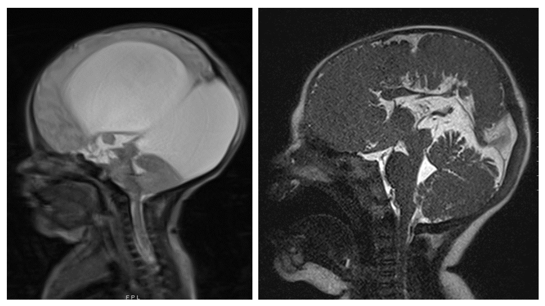
Although prenatal ultrasound is an excellent tool, fetal magnetic resonance imaging (MRI) is often used as an adjunct to obtain a more complete characterization of an encephalocele. Fetal MRI may be particularly useful when the sonographic views are limited by maternal body habitus, fetal position, or oligohydramnios. In addition to confirming the presence of a skull defect, the volume and gyral pattern of the herniated brain parenchyma can be evaluated (Figure 1) (8, 13, 23). Other cerebral malformations can also be better assessed with fetal MRI, and the spine can be evaluated for the presence of a spinal dysraphism and/or syringomyelia.
 

Prenatal MRI of a patient with encephalocele


Historical case series have reported that approximately 50% of fetuses with encephaloceles die prior to birth (5, 24-26). An accurate understanding of the prenatal natural history of encephaloceles is complicated by the high rate of termination of affected pregnancies. However, progressive herniation over the course of the pregnancy has been described (27). In particular, a recent study of 20 patients with fetal and postnatal MRIs (9 of whom had a meningocele and 11 of whom had an encephalocele, identified progressive herniation in 50% of the cases. Progressive herniation was defined as either an absolute increase of 5% or more in the volume of neural tissue within the fetal cephalocele, or as herniation of a new structure into the cephalocele sac (5). The authors found that prenatal hindbrain herniation and/or prenatal microcephaly are associated with a risk of progressive cephalocele herniation.

Prenatal counseling should take place as early as possible so that parents may make an informed decision to continue versus terminate the pregnancy. Cell-free DNA screening, amniocentesis, or chorionic villus sampling can be performed to detect genetic syndromes and chromosomal abnormalities, and facilitates genetic counselling. The pregnancy is otherwise managed according to standard obstetric guidelines.

In an effort to prevent the progressive herniation of brain parenchyma into the cephalocele sac over the course of a pregnancy, Cavalheiro et al. performed fetal repairs of 9 encephaloceles (28). Only those cases in which the amount of herniated brain parenchyma was less than 20% of the total volume of the cephalocele sac were offered fetal repair. The surgical technique involved exposure of the encephalocele through a hysterotomy, incision of the herniated sac, exposure and circumferential dissection of the meningeal tissue, excision versus internal relocation of the neural tissue, closure of the meningeal layer and dura, placement of an absorbable miniplate to reconstruct the bony defect, and closure of the skin. The authors reported that fetal repair reversed the trend toward microcephaly and led to improved neurodevelopmental outcomes, with only 2 of 9 patients exhibiting developmental delays on the Bayley Scales of Infant Development II.

Others have pointed out that although this study demonstrates the feasibility of fetal encephalocele repair, it remains unclear whether fetal repair results in better outcomes than postnatal repair (29). Proponents of fetal encephalocele repair compare it to fetal myelomeningocele repair, the efficacy of which has been well established (30). However, cephaloceles differ in that they are closed, skin-covered neural tube defects. Unlike myelomeningoceles, the neural tissue in encephaloceles is not progressively damaged by exposure to the amniotic fluid – therefore the same rationale cannot be used to justify fetal repair. On the other hand, fetal repair might prevent the progressive herniation of brain parenchyma over the course of the pregnancy. Would this result in better outcomes? Although several studies have indicated that a large amount of neural tissue within the cephalocele is a negative prognostic indicator (25, 31), more recent studies suggest that migrational anomalies detected on MRI are more predictive of neurocognitive outcomes than the volume of herniated brain tissue (4). To justify the maternal and fetal risks of prenatal repair, the prognostic significance of preventing progressive herniation into the cephalocele sac needs to be established.
 

The delivery should take place in a hospital with immediate access to maternal-fetal-medicine specialists, neonatal intensivists, and pediatric neurosurgeons. Many obstetricians prefer a planned Caesarean section to avoid trauma to the herniated neural tissue and to facilitate coordination of the various teams that will be involved immediately after the delivery. Caesarean sections are certainly recommended for large encephaloceles, though vaginal delivery can be considered in select cases for small encephaloceles.

The initial assessment should focus on the airway, breathing, and circulation of the neonate. Once stabilized, a full physical examination, including a complete neurological assessment (cranial nerves, motor strength, tone, sensation, and reflexes) should be performed. The cephalocele sac should be assessed, and in particular the integrity of the skin overlying the sac should be evaluated to determine the risk of a cerebrospinal fluid (CSF) leak.

A postnatal MRI should be obtained to fully evaluate the cranial defect and characterize the herniated brain parenchyma, if any, that is present in the sac (Figure 2). MRI can also be used to identify other intracranial anomalies, such as ventriculomegaly, a Chiari malformation, Dandy-Walker malformation, holoprosencephaly, or agenesis of the corpus callosum. In addition to standard MRI sequences, vascular imaging (i.e. magnetic resonance angiography and venography) should be obtained to determine the presence of vascular structures within the sac, as well as to characterize the relationship between the cephalocele and the dural venous sinuses (8). 

Postnatal MRI of a patient with encephalocele


Multidisciplinary postnatal care is essential. The team often includes a neurosurgeon, neurologist, intensivist, geneticist, craniofacial specialist, and an endocrinologist. An endocrinological evaluation is necessary, as endocrine abnormalities are frequently identified among patients with congenital malformations involving midline cranial structures.

Surgical repair is the primary treatment for an encephalocele. The goals of surgery are to remove non-functional neural tissue, preserve functional tissue, obtain a watertight dural closure, reconstruct the bony defect (when it is large), and remove excess skin (4, 8). The surgical repair of an occipital encephalocele can typically be performed electively after several months of age, unless the skin is thin, ulcerated, and at risk of leaking CSF, in which case urgent surgery should be performed to prevent meningitis. The presence of hydrocephalus and the impact of the size of the sac on the ability to position and care for the neonate are other considerations that may impact the timing of surgery. For sincipital and basal encephaloceles, too, the risks of blood loss must be balanced with the risks of progressive facial deformity, airway compromise, and infection. In the absence of rhinorrhea, surgery is often delayed until 2-3 years of age, though some have recommended early surgery if there is sufficient nasal expansion (32).

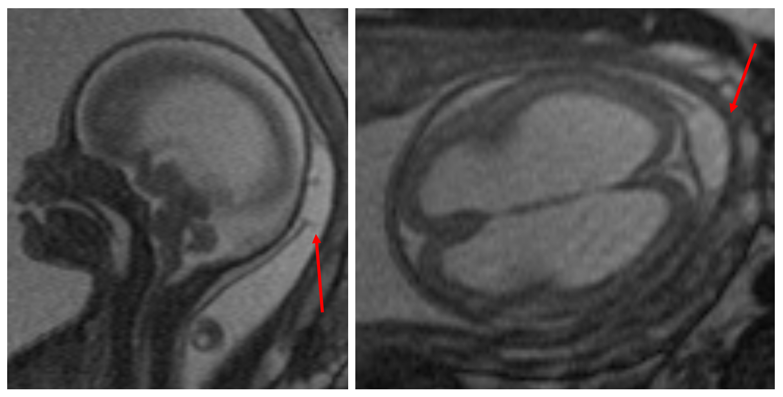
The patient with an occipital encephalocele must be carefully intubated, either in the supine position on a donut or horseshoe head holder, or in a lateral position. The patient is then placed in the prone position, taking care to pad all bony prominences and avoid pressure on the eyes (Figure 3). 

Prep for surgical intervention for a patient with encephalocele


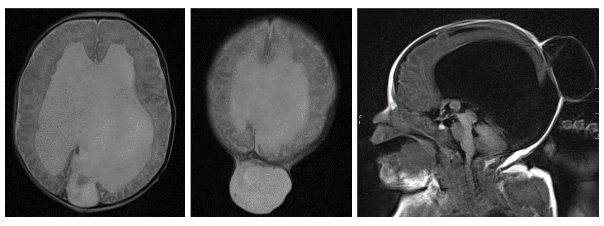
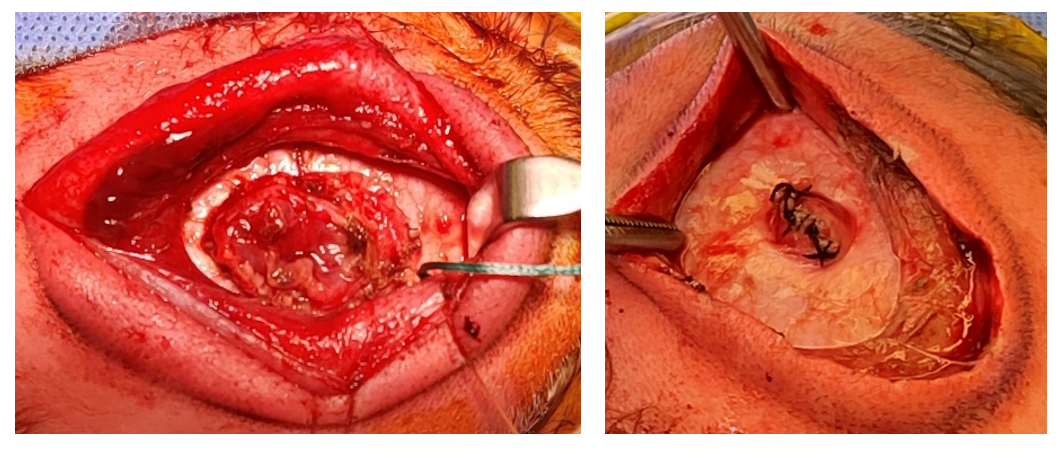
After the occipital region is sterilely prepped and draped, and preoperative antibiotics are administered, either a linear or elliptical incision is made in the skin of the encephalocele sac. The dissection is carried down to the pericranium and the bony edges are defined. Non-functional neural tissue is then coagulated and resected, while tissue that is suspected to be functional is preserved and restored to the intracranial space. Excess dura is excised, and a primary, watertight dural closure is performed (Figure 4). When a large amount of functional tissue is protruding into the cephalocele sac, the dura is closed around the herniated tissue and a cranial vault expansion can be performed, either in the same setting or a second stage procedure, to create space for the neural tissue. Autologous split thickness calvarial grafts can be used to reconstruct the bony defect when it is large; alternatively, small defects will often heal spontaneously (33). 

Watertight Dural Closure of Encephalocele

When hydrocephalus is present preoperatively, shunt placement is often performed either 
before or during the encephalocele repair. Alternatively, an external ventricular drain may be placed at the time of the initial repair. When CSF diversion is not performed, close postoperative monitoring is mandatory to assess for the development of worsening hydrocephalus postoperatively. The head circumference should be tracked, serial imaging should be performed to evaluate the size of the ventricles, and the incision should be monitored for pseudomeningocele formation or CSF leakage. There should be a low threshold for shunt placement when indicated (Figure 5) (33). Endoscopic third ventriculostomy has also been described as a treatment option for encephalocele-associated hydrocephalus (34-36). 
 

Post-op imaging of encephalocele patient

Survival

Mortality rates vary widely in the literature. Older series reported that in addition to the 50% of fetuses who die in utero, another one-third of infants born with an encephalocele die, typically within the 1st year of life (24-26). More recent studies, however, have reported lower mortality rates of approximately 5% (2, 4, 31). The discrepancy may be due, in part, to differences in the proportion of posterior versus anterior encephaloceles, the size of the encephalocele, and the amount of herniated brain tissue. Low birth weight and the presence of multiple intracranial anomalies are associated with an increased risk of mortality (26).


Hydrocephalus

Approximately 60-90% of posterior encephaloceles and 10-15% of anterior encephaloceles are associated with hydrocephalus (2, 31, 37, 38). Among occipital encephaloceles, supratorcular encephaloceles have the highest rate of hydrocephalus (60%) followed by the infratorcular subtype (47%). Torcular encephaloceles have a lower rate of hydrocephalus (25%), which is suspected to be a result of the high mortality rate associated with this subtype (39). Hydrocephalus may be present at birth, or may develop following encephalocele repair (2, 31, 40). Some authors have hypothesized that progressive hydrocephalus following the surgical repair may result from torsion of the cerebral aqueduct or the venous sinuses, or alternatively may be related to postoperative meningitis (34, 36, 40, 41). A third possibility is that the encephalocele sac acts as a “5th ventricle” and once that space is eliminated, CSF is re-routed and leads to progressive ventriculomegaly (41). Regardless of the underlying etiology, Da Silva et al. found that the presence of neural tissue within the sac was the only statistically significant prognostic factor for hydrocephalus (31), while Kankam et al. found that a larger encephalocele sac, the presence of other associated anomalies, and infections were significant independent predictors of hydrocephalus (41). Early shunt placement prior to or during the encephalocele repair has been advocated by some to facilitate the closure and minimize the risk of a CSF leak (2, 40, 42, 43), while others recommend shunt placement in a second-stage procedure following the repair to minimize the risk of infection (39). If the latter approach is taken, patients must be monitored closely for progressive ventriculomegaly and symptomatic hydrocephalus post-repair (41).


Development

Neurodevelopmental outcomes vary widely across studies. Some studies have reported overall rates of neurodevelopmental delay ranging from 16%-33% (2, 44, 45), while others have identified overall rates in the range of 50% - 60% (4, 31).

Lo et al. studied the outcomes of 68 encephaloceles and 17 meningoceles treated at the Hospital for Sick Children between 1990 and 2006 (4). Though the overall mortality rate was only 5%, there was a high rate of developmental delay among survivors – 48% had normal development, 11% had mild delays, 16% had moderate delays, and 25% had severe delays. The location of the cephalocele was not found to be predictive of outcome; a multivariable analysis demonstrated that hydrocephalus (OR 10.9, 95% CI 2.1 – 55.9) and the presence of intracranial anomalies (OR 4.1, 95% CI 1.3 – 12.6) were significant predictors of developmental delay.

Da Silva et al. studied 70 patients with encephaloceles that were treated at Children’s Hospital Los Angeles between 1994 and 2012 (31). Overall, 40% had a normal neurological outcome, 40% had a mild or moderate delay, and 20% had a severe delay. A multivariable analysis found that the only significant prognostic factor was the presence of hydrocephalus (OR 17.2, 95% CI 1.7 – infinity).

Kankam et al. studied 102 patients with cephaloceles (42% of which contained neural tissue) that were treated at Children’s Medical Center Hospital in Tehran between 2010 and 2021 (2). The authors screened for developmental milestones that were categorized into 4 domains, including social and emotional development, language/communication, cognitive, and movement/physical development. Patients were classified as having a neurodevelopmental delay if there were delays in 2 or more domains. Of the 94 patients with complete developmental evaluations, 67% had normal neurodevelopment, 15% had mild or moderate delays, and 18% had severe delays. A multivariable analysis demonstrated that the presence of neural tissue in the encephalocele sac was the only predictor of neurodevelopmental delay (OR 7.0, 95% CI 1.3 – 37.2).

Encephalocele development is suspected to be multifactorial, involving both genetic and environmental factors. Unlike open neural tube defects involving the spine, folate has not been definitively implicated in the pathogenesis of encephaloceles. Encephaloceles have been associated with trisomy 18, as well as with more than 30 syndromes, including Meckel-Gruber syndrome, amniotic band syndrome, Knobloch syndrome, Walker-Warburg syndrome, Fraser syndrome, and Roberts syndrome (8, 13).

The genetic syndrome most closely associated with encephaloceles is Meckel-Gruber syndrome, an autosomal recessive condition characterized by the triad of an occipital cephalocele, axial polydactyly, and polycystic kidneys (22, 46). The syndrome belongs to a group of ciliopathies, most often caused by a mutation of the MKS1 gene (47). Sonographic evaluation of the fetal kidneys during the first trimester is often helpful in establishing the diagnosis (20).

Genetic testing and routine evaluation of the karyotype should be performed for all patients with an encephalocele (48). Genetic abnormalities have been identified in up to 14% of affected fetuses when routine karyotyping was performed; even higher rates of genetic abnormalities were identified with microarray testing and whole exome sequencing (47, 49, 50). The risk of recurrence is estimated to be 3-5% in the setting of an isolated encephalocele, and 25% in the setting of an autosomal recessive condition.

  1. Naidich TP, Altman NR, Braffman BH, McLone DG, Zimmerman RA. Cephaloceles and related malformations. AJNR Am J Neuroradiol. 1992;13(2):655-690.
  2. Kankam SB, Tavallaii A, Mohammadi E, Nejat A, Habibi Z, Nejat F. The neurodevelopmental outcomes of children with encephalocele: a series of 102 patients. J Neurosurg Pediatr. 2023;31(2):151-158.
  3. Vakharia VN, Toescu S, Copp AJ, Thompson DNP. A topographical analysis of encephalocele locations: generation of a standardised atlas and cluster analysis. Childs Nerv Syst. 2023.
  4. Lo BW, Kulkarni AV, Rutka JT, et al. Clinical predictors of developmental outcome in patients with cephaloceles. J Neurosurg Pediatr. 2008;2(4):254-257.
  5. Gadgil N, McClugage SG, Aldave G, et al. Natural history of posterior fetal cephaloceles and incidence of progressive cephalocele herniation. J Neurosurg Pediatr. 2022:1-7.
  6. Parker SE, Mai CT, Canfield MA, et al. Updated National Birth Prevalence estimates for selected birth defects in the United States, 2004-2006. Birth Defects Res A Clin Mol Teratol. 2010;88(12):1008-1016.
  7. Rowland CA, Correa A, Cragan JD, Alverson CJ. Are encephaloceles neural tube defects? Pediatrics. 2006;118(3):916-923.
  8. Markovic I, Bosnjakovic P, Milenkovic Z. Occipital Encephalocele: Cause, Incidence, Neuroimaging and Surgical Management. Curr Pediatr Rev. 2020;16(3):200-205.
  9. Macfarlane R, Rutka JT, Armstrong D, et al. Encephaloceles of the anterior cranial fossa. Pediatr Neurosurg. 1995;23(3):148-158.
  10. Mahapatra AK, Agrawal D. Anterior encephaloceles: a series of 103 cases over 32 years. J Clin Neurosci. 2006;13(5):536-539.
  11. Achar SV, Dutta HK. Sincipital Encephaloceles: A Study of Associated Brain Malformations. J Clin Imaging Sci. 2016;6:20.
  12. Hunt JA, Hobar PC. Common craniofacial anomalies: facial clefts and encephaloceles. Plast Reconstr Surg. 2003;112(2):606-615; quiz 616,722.
  13. Weichert J, Hoellen F, Krapp M, et al. Fetal cephaloceles: prenatal diagnosis and course of pregnancy in 65 consecutive cases. Arch Gynecol Obstet. 2017;296(3):455-463.
  14. Cameron M, Moran P. Prenatal screening and diagnosis of neural tube defects. Prenat Diagn. 2009;29(4):402-411.
  15. Blaas HG, Eik-Nes SH. Sonoembryology and early prenatal diagnosis of neural anomalies. Prenat Diagn. 2009;29(4):312-325.
  16. Miskin M, Rudd NL, Dische MR, Benzie R, Pirani BB. Prenatal ultrasonic diagnosis of occipital encephalocele. Am J Obstet Gynecol. 1978;130(5):585-587.
  17. Budorick NE, Pretorius DH, McGahan JP, Grafe MR, James HE, Slivka J. Cephalocele detection in utero: sonographic and clinical features. Ultrasound Obstet Gynecol. 1995;5(2):77-85.
  18. Graham D, Johnson TR, Jr., Winn K, Sanders RC. The role of sonography in the prenatal diagnosis and management of encephalocele. J Ultrasound Med. 1982;1(3):111-115.
  19. Goldstein RB, LaPidus AS, Filly RA. Fetal cephaloceles: diagnosis with US. Radiology. 1991;180(3):803-808.
  20. Sepulveda W, Wong AE, Andreeva E, Odegova N, Martinez-Ten P, Meagher S. Sonographic spectrum of first-trimester fetal cephalocele: review of 35 cases. Ultrasound Obstet Gynecol. 2015;46(1):29-33.
  21. Ong AGJ, Rolnik DL, Menezes M, Meagher S. Early Diagnosis and Differences in Progression of Fetal Encephalocele. J Ultrasound Med. 2020;39(7):1435-1440.
  22. Sepulveda W, Sebire NJ, Souka A, Snijders RJ, Nicolaides KH. Diagnosis of the Meckel-Gruber syndrome at eleven to fourteen weeks' gestation. Am J Obstet Gynecol. 1997;176(2):316-319.
  23. Kasprian GJ, Paldino MJ, Mehollin-Ray AR, et al. Prenatal imaging of occipital encephaloceles. Fetal Diagn Ther. 2015;37(3):241-248.
  24. Mealey J, Jr., Dzenitis AJ, Hockey AA. The prognosis of encephaloceles. J Neurosurg. 1970;32(2):209-218.
  25. Kiymaz N, Yilmaz N, Demir I, Keskin S. Prognostic factors in patients with occipital encephalocele. Pediatr Neurosurg. 2010;46(1):6-11.
  26. Siffel C, Wong LY, Olney RS, Correa A. Survival of infants diagnosed with encephalocele in Atlanta, 1979-98. Paediatr Perinat Epidemiol. 2003;17(1):40-48.
  27. Gandhoke GS, Goldschmidt E, Kellogg R, Greene S. Encephalocele development from a congenital meningocele: case report. J Neurosurg Pediatr. 2017;20(5):419-422.
  28. Cavalheiro S, Silva da Costa MD, Nicacio JM, et al. Fetal surgery for occipital encephalocele. J Neurosurg Pediatr. 2020;26(6):605-612.
  29. Gupta N. Editorial. Fetal repair of encephaloceles. J Neurosurg Pediatr. 2020;26(6):603-604.
  30. Adzick NS, Thom EA, Spong CY, et al. A randomized trial of prenatal versus postnatal repair of myelomeningocele. N Engl J Med. 2011;364(11):993-1004.
  31. Da Silva SL, Jeelani Y, Dang H, Krieger MD, McComb JG. Risk factors for hydrocephalus and neurological deficit in children born with an encephalocele. J Neurosurg Pediatr. 2015;15(4):392-398.
  32. Thompson HM, Schlosser RJ, McCarty Walsh E, et al. Current management of congenital anterior cranial base encephaloceles. Int J Pediatr Otorhinolaryngol. 2020;131:109868.
  33. Selden N, Baird L. Neurosurgery by Example: Pediatric Neurosurgery. Oxford University Press; 2019.
  34. Munekata Y, Sugiyama T, Ueda Y, Mizushima M, Motegi H, Fujimura M. Late-onset obstructive hydrocephalus associated with occipital encephalocele with large skull defect successfully treated by endoscopic third ventriculostomy. Childs Nerv Syst. 2023;39(1):307-310.
  35. Moorthy RK, Rajshekhar V. Management of hydrocephalus associated with occipital encephalocoele using endoscopic third ventriculostomy: report of two cases. Surg Neurol. 2002;57(5):351-355; discussion 355.
  36. Warf BC, Stagno V, Mugamba J. Encephalocele in Uganda: ethnic distinctions in lesion location, endoscopic management of hydrocephalus, and survival in 110 consecutive children. J Neurosurg Pediatr. 2011;7(1):88-93.
  37. Gamache FW, Jr. Treatment of hydrocephalus in patients with meningomyelocele or encephalocele: a recent series. Childs Nerv Syst. 1995;11(8):487-488.
  38. Bui CJ, Tubbs RS, Shannon CN, et al. Institutional experience with cranial vault encephaloceles. J Neurosurg. 2007;107(1 Suppl):22-25.
  39. Protzenko T, Dos Santos Gomes Junior SC, Bellas A, Salomao JFM. Hydrocephalus and occipital encephaloceles: presentation of a series and review of the literature. Childs Nerv Syst. 2021;37(11):3437-3445.
  40. Refaee EAE, Refaat MI, Reda M. Incidence of Secondary Hydrocephalus after Excision of Huge Encephaloceles in Neonates: Case Study. J Neurol Surg A Cent Eur Neurosurg. 2018;79(1):15-18.
  41. Kankam SB, Nejat A, Tavallaii A, Tayebi Meybodi K, Habibi Z, Nejat F. Hydrocephalus in patients with encephalocele: introduction of a scoring system for estimating the likelihood of hydrocephalus based on an 11-year experience from a tertiary center. J Neurosurg Pediatr. 2023;31(4):298-305.
  42. Rehman L, Farooq G, Bukhari I. Neurosurgical Interventions for Occipital Encephalocele. Asian J Neurosurg. 2018;13(2):233-237.
  43. Kabre A, Zabsonre DS, Sanou A, Bako Y. The cephaloceles: A clinical, epidemiological and therapeutic study of 50 cases. Neurochirurgie. 2015;61(4):250-254.
  44. Date I, Yagyu Y, Asari S, Ohmoto T. Long-term outcome in surgically treated encephalocele. Surg Neurol. 1993;40(2):125-130.
  45. Martinez-Lage JF, Poza M, Sola J, et al. The child with a cephalocele: etiology, neuroimaging, and outcome. Childs Nerv Syst. 1996;12(9):540-550.
  46. Alexiev BA, Lin X, Sun CC, Brenner DS. Meckel-Gruber syndrome: pathologic manifestations, minimal diagnostic criteria, and differential diagnosis. Arch Pathol Lab Med. 2006;130(8):1236-1238.
  47. Dabkowska S, Kucinska-Chahwan A, Beneturska A, et al. Prenatal diagnosis and clinical significance of cephalocele-A single institution experience and literature review. Prenat Diagn. 2020;40(5):612-617.
  48. Practice Bulletin No. 187 Summary: Neural Tube Defects. Obstet Gynecol. 2017;130(6):1394-1396.
  49. Wininger SJ, Donnenfeld AE. Syndromes identified in fetuses with prenatally diagnosed cephaloceles. Prenat Diagn. 1994;14(9):839-843.
  50. Sepulveda W, Corral E, Ayala C, Be C, Gutierrez J, Vasquez P. Chromosomal abnormalities in fetuses with open neural tube defects: prenatal identification with ultrasound. Ultrasound Obstet Gynecol. 2004;23(4):352-356.