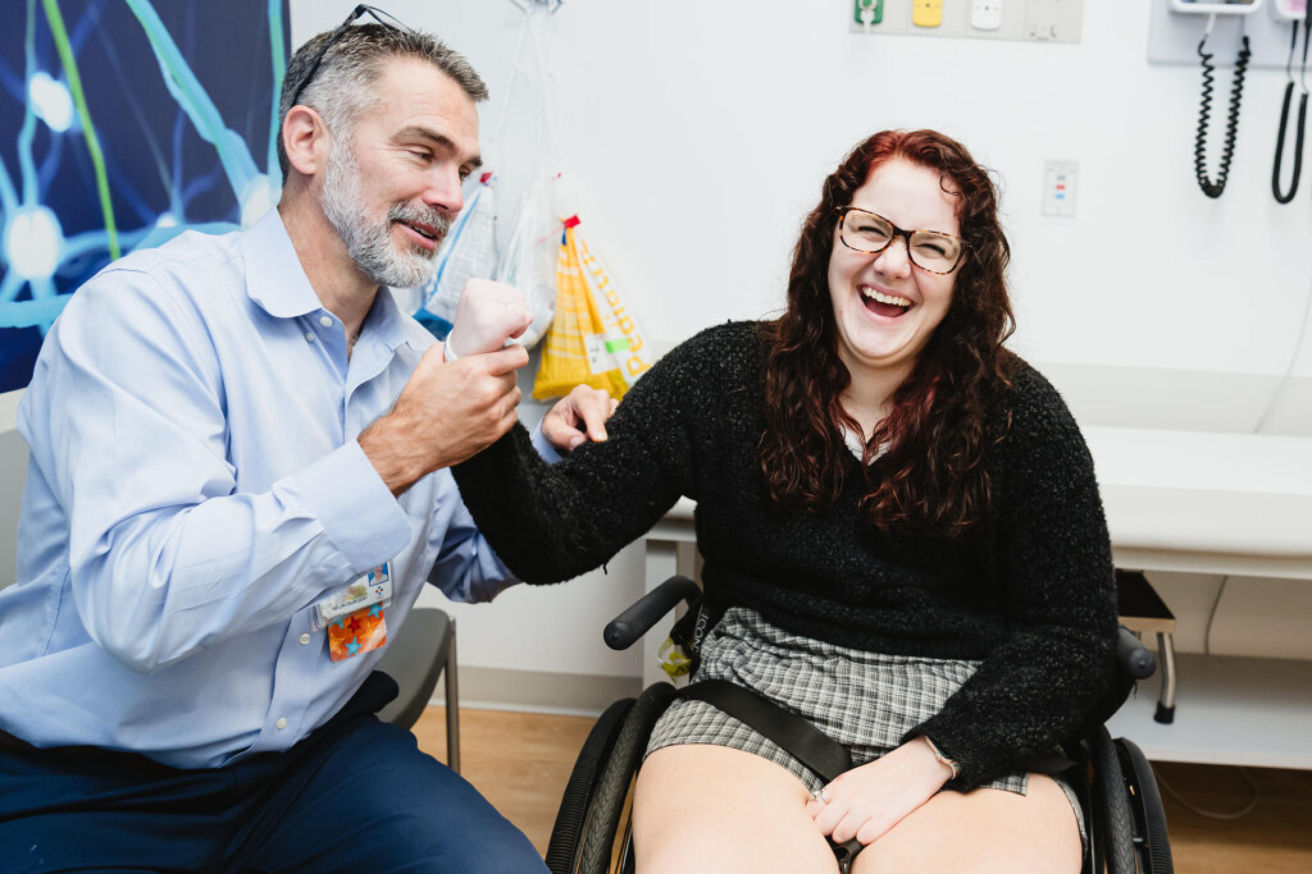
Neurosurgery Conditions & Treatments Brain and Spinal Cord Tumors Central nervous system tumors involving the brain or spinal cord represent the largest category of solid tumors afflicting children. Roughly four in every 100,000 children in North America will have a brain tumor. They are rarely related to syndromes or inherited mutations, with most brain tumors arising by chance due to errors in cell division that cannot be passed on to future generations. Most pediatric nervous system tumors arise from support cells within the brain or spinal cord called glial cells, but tumors can form from virtually any cell type or its precursor – including the neurons themselves, the coverings of the brain and spinal cord, the bones of the skull, the cells that secrete spinal fluid, and the cells that regulate hormones throughout the body. Most Common Pediatric Brain Tumors Tumor TypeSynopsisTreatment GliomaTumors arising from the support cells of the brain—glial cells or astrocytes. This group includes juvenile pilocytic astrocytomas, and they comprise approximately 60% of all pediatric brain tumors. Most will be benign, though malignant variants exist.Surgical resection, if deemed safe, is the mainstay of therapy for gliomas. In the case of juvenile pilocytic astrocytomas and other low-grade gliomas, surgery alone may be sufficient to provide a cure. For patients who cannot be cured with surgery and who have evidence of progressive disease, targeted chemotherapy and/or radiation therapy may be required. MedulloblastomaPart of the family of tumors known as primitive neuroectodermal tumors (PNETs), this is the most common malignant tumor in children. They account for approximately 30% of all pediatric brain tumors.Maximal, safe surgical resection is an essential part of any medulloblastoma treatment plan. Complete cure with surgery alone is generally not possible, though, and chemotherapy and radiation is often required, with the exact regimen dictated by the molecular subtype of the tumor. Ependymomaline the inner surfaces of the brain and spinal cord, where spinal fluid circulates. These tumors account for approximately 10% of all pediatric brain tumors, and they have a propensity for spreading throughout the head and spine by shedding cells into the spinal fluid.Maximal, safe surgical resection is the primary treatment modality for ependymomas. Chemotherapy and radiation therapy are also employed following surgery to eradicate microscopic disease that may be left after all visible tumor has been removed. Presentation Because of the remarkable plasticity of the developing nervous system, children with brain and spinal cord tumors will often present with much more subtle symptoms than adults with similar tumors. Early symptoms of a central nervous system tumor can include nausea, vision loss, rapid head growth, balance difficulties, difficulty learning to walk, or persistent head tilt. When a child’s tumor overwhelms their capacity to compensate for the mass, they will often come to medical attention with more dramatic findings, such as severe headache and vomiting. Symptoms of Brain and Spinal Cord Tumors Tumor LocationPotential Symptons Cerebrum• Weakness • Numbness • Difficulty with speech • Seizures • Headaches • Nausea/vomiting Brainstem or Cerebellum• Headaches • Nausea/vomiting • Weakness • Difficulty with balance • Difficulty with walking • Double vision • Problems swallowing Spinal Cord• Difficulty with walking • Weakness • Problems controlling bladder • Numbness • Deformities of the back and legs • Pain in the neck, back and legs Diagnosis A medical professional will use a variety of clinical, radiographic and laboratory tests to try to establish whether a child truly has a nervous system tumor or not. The most important component of any evaluation, however, is the clinical history and physical exam that your child’s neuro-specialist takes. Possible Diagnostics ModalityTestsInformation Provided Radiography• MRI of the brain and/or spine • CT scan of the brain, spine or (rarely) body • X-rays • PET scan Imaging of the head and spine provides important information about the extent of the patient’s disease and what critical structures might be affected by further attempts to diagnose or treat the tumor. In most cases, imaging will include the entire head and spine, as many brain tumors have the potential to spread to the spine. A definitive diagnosis by imaging alone is often not possible, and a tissue sample is almost always required for a definitive diagnosis. Laboratory• MRI of the brain and/or spine • CT scan of the brain, spine or (rarely) body • X-rays • PET scan • Tumor markers (ex. CEA, AFP, βHCG, lactate) • Hormone levels (ex. TSH, TH, GH, ACTH, cortisol, prolactin) can be helpful in the early diagnosis or staging of some pediatric tumors, particularly pituitary or pineal region tumors. In specific cases, bloodwork or spinal fluid tests can establish a diagnosis without the need for a tissue biopsy. Procedures• Angiogram • Lumbar puncture • Stereotactic biopsy Lumbar puncture and stereotactic biopsy may be part of the workup for some tumors, particularly when it is advantageous to have a definitive diagnosis prior to considering an attempt at surgical resection. Angiograms – a procedure that allows for high-resolution images of a patient’s blood vessels to be taken – can also provide valuable information about tumors with a high propensity for bleeding or located near critical vessels. In certain cases, an angiogram may also allow for vascular tumors to be partially treated prior to attempting a conventional surgical resection. Pathology• Cytology • Histopathology • Genetic / molecular typing Ultimately, only a pathologist review of a sample from a tumor can definitively provide a diagnosis. Molecular and genetic testing of the tumor also provides information that can predict how a tumor is likely to behave in the future and which types of therapy will best treat it. Treatment Pediatric brain and spinal tumor treatments are devised in a multidisciplinary fashion. Each child’s case is reviewed by neurosurgery, neuro-oncology, neuro-radiology, and radiation oncology. When appropriate, additional specialties such as neurology, ophthalmology, endocrinology or general surgery may need to be involved, such as when a patient’s tumor is suspected to cause dysfunctions particular to other organs systems in the body. In most cases, the initial step in treating a nervous system tumor will be to attempt surgical removal of the tumor. Our team of neurosurgeons has extensive experience safely removing some the most challenging types of brain and spinal cord tumors in pediatric neuro-oncology. Your surgeon and the neuro-oncology team will guide you, step by step, through your child’s surgical plan and expected post-operative recovery. We use advanced stereotactic, neuro-endoscopic, and minimally invasive techniques, wherever possible, to minimize the potential surgical risks to our patients while accelerating patient recovery. If chemotherapy or radiation therapy are required, our team of neuro-oncologists and radiation oncologists will tailor the therapeutic regimen your child receives to the particular molecular profile of their tumor. In cases where pediatric nervous system tumors possess exploitable mutations, this approach can produce improved tumor control and limited treatment-related side effects. Long-Term Treatment We strive to ensure that all our pediatric tumor patients live long and active lives. In pursuit of that aim, our tumor care team follows patients out into young adulthood to watch for possible late effects of cancer treatments. Our team of oncologists, physical therapists, audiologists, cardiologists, psychologists and endocrinologists continue to monitor and support our patients throughout their developing years so that we can provide them with the best footing for transitioning to an adulthood beyond their childhood cancer diagnosis. Craniosynostosis Craniosynostosis is a condition whereby one or more of the bony plates in a child’s skull fuse prematurely and impair normal cranial growth. This condition occurs in roughly one in 2,000 children, and is typically identified within the first few months of life. Left untreated, children with craniosynostosis will go on to have lifelong deformities of their head and, in severe cases, visual problems, headaches and vomiting. Most craniosynostosis occurs in an isolated manner, without the child suffering any additional congenital deformities of the face or limbs. Hereditary craniosynostosis does occur, but it is the exception rather than the rule; individuals born with isolated craniosynostosis are unlikely to have children with the same problem. About 20% of the time, craniosynostosis occurs as one aspect of a broader craniofacial syndrome such as Aperts or Crouzon, and these types of syndromic craniosynostosis are often more prone to have multiple bony fusions and increased pressure within the head. Syndromic craniosynostosis patients will often require the assistance of multiple pediatric specialists working to address extremity, mouth, nose, eye, ear and cranial abnormalities at the same time. The mechanism by which craniosynostosis arises is still not well understood, but some genetic mutations have been suggested in laboratory models. Diagnosis Often, the diagnosis of craniosynostosis can be made during an initial evaluation with a craniofacial specialist. An expert craniofacial surgeon can diagnose most forms of craniosynostosis based on the appearance of the child’s cranial deformity, as each type of craniosynostosis produces stereotypical changes in the shape of the skull. For more complicated cases or for syndromic patients, more detailed radiographic studies may be useful for both diagnostic and surgical planning purposes. The most important distinction your child’s physician will make is between craniosynostosis and other, benign types of cranial deformity. Up to 50% of newborns will have some type of cranial deformity within the first few months of life, but most of these deformities are due to molding in the womb or from lying on the back of the head. Cranial deformities that result from external molding of the child’s head are broadly classified as plagiocephaly: These patients improve without the need for surgery. We use a number of methods to diagnose cranial deformities, including our highly experienced craniofacial surgeons, in-office cranial measurements and same-day ultrasounds. In most cases, an accurate diagnosis can be made in one visit and without the need to expose patients to radiation or sedate them for lengthy radiographic studies. Diagnostic Studies StudyDescription COMPUTED TOMOGRAPHY (CT)• Detailed 3D images of the skull • Most useful for complex or syndromic patients • Uses radiation to create an image • Usually not necessary for simple, non-syndromic cases MAGNETIC RESONANCE IMAGING (MRI)• Best for imaging the brain • Can create 3D images of the skull comparable to CT • Uses magnetic waves to create an image • Usually requires that the patient be sedated for the study X-RAY• Quick • Can usually assess whether most of the major cranial sutures are open • Not always possible to visualize all the cranial sutures ULTRASOUND• Quick • Can usually assess whether most of the major cranial sutures are open • No radiation or sedation • Not accurate when examining the cranial suture in the middle of the forehead Treatment Very mild cases of craniosynostosis may not require surgery, but the majority of craniosynostosis patients will require surgical correction to address their deformity and return skull growth to normal. Surgical outcomes tend to be much better the earlier in life the patient receives surgical attention, likely due to the increased plasticity of the infant skull before 1 year of life. Early identification of craniosynostosis is the first step in treatment. Surgery to correct craniosynostosis can be performed in several ways, depending on the age of the patient and the type of deformity. No single method has been shown to produce superior cosmetic or functional outcomes, and so our philosophy is to always employ the least invasive surgical technique appropriate for that child’s deformity. Children under 6 months of age often have a minimally invasive, endoscopic surgery. Older children will more commonly benefit from an open cranial reconstruction, performed by our joint plastic surgery and neurosurgery craniofacial team. But age alone is not the only factor in determining who is eligible for either procedure: Your craniofacial surgeon will consider your child’s entire medical condition before making their recommendation. Long-Term Care Late complications after surgical repair of craniosynostosis are rare, but your child will require continued monitoring for several years after surgery to look for late impacts to vision, secondary fusions of the skull, persistent skull defects, and trends in their neurodevelopment. Patients whose craniosynostosis was part of an overarching syndrome may require additional screening or treatment for other abnormalities of the face and ears as well. Our multidisciplinary Craniofacial Clinic allows families to be seen by expert pediatric and surgical specialists from all craniofacial disciplines in a single location, providing comprehensive and convenient pre- and post-surgical care from birth to young adulthood. Cerebal palsy and spasticity Spasticity is a condition of increased muscle tone that is commonly seen in Cerebral palsy. As a result of increased muscle tone, children with spasticity may experience pain or musculoskeletal deformity. In many cases, children receive care from neurologists, physical and occupational therapists, physiatrists, and orthopedic surgeons to improve their comfort and mobility. Connecticut Children’s Division of Neurosurgery offers surgical treatments to treat spasticity in selected patients. These treatment options include: Intrathecal baclofen pump placement: A pump system placed under the skin delivers relaxing medication directly into the spinal fluid surrounding the spinal cord Selective dorsal rhizotomy: Selected nerve roots in the lower back are sectioned to reduce the action of overactive muscles in the legs Our pediatric specialists will review these options at your child’s visit. Chiari Malformation Chiari malformation is a condition in which brain tissue extends into the spinal canal – simply put, the brain is too big for the head. It occurs when part of a child’s skull is abnormally small or misshapen, pressing on the brain and forcing it downward. Current estimates are that up to 1% of children have this malformation. Doctors categorize Chiari malformation into several types depending on the anatomy of the brain tissue displaced into the spinal canal and whether developmental abnormalities of the brain or spine are present. Children with Chiari malformation can experience symptoms related to irritation of the covering of the brain, direct compression of brain structures, or dilation of the spinal cord due to an abnormal flow of spinal fluid (syringomyelia). Symptoms can include: Headaches, often with coughing, sneezing or straining Neck pain Unsteady gait (problems with balance) Poor hand coordination (fine motor skills) Numbness and tingling of the hands and feet Dizziness Difficulty swallowing, sometimes accompanied by gagging, choking and vomiting Speech problems, such as hoarseness Less often, children with Chiari malformation may experience: Ringing or buzzing in the ears (tinnitus) Weakness Slow heart rhythm Curvature of the spine (scoliosis) related to spinal cord impairment Abnormal breathing, such as central sleep apnea (in which a child stops breathing during sleep) Treatment of Chiari malformation – depending on the type, severity and associated symptoms – can include regular monitoring, medications and surgery. In some cases, no treatment is needed. Connecticut Children’s team of pediatric neurosurgery specialists are experts in diagnosing and treating Chiari malformation. We’ll review the options with you and help you select the best treatment for your child. Hydrocephalus Hydrocephalus is a condition that occurs when cerebrospinal fluid (CSF) builds up in the brain, typically due to an imbalance between the production and absorption of cerebral spinal fluid. This often results in increased intracranial pressure, which can cause long-term issues if left untreated. Hydrocephalus can occur on its own or be associated with other conditions. Diagnosing Hydrocephalus Hydrocephalus may present with a variety of signs and symptoms: Infants Head enlargement Tense or bulging soft spot Prominent scalp veins Persistent vomiting Less interest in feeding Irritability and/or sleepiness Downward deviation of the eyes Children, Adolescents and Adults Persistent vomiting Persistent headache Irritability and/or sleepiness Downward deviation of the eyes If your child is displaying any signs or symptoms of hydrocephalus, imaging may be necessary to evaluate the fluid in their brain. Hydrocephalus can be diagnosed using three major techniques: Ultrasound (US) Computerized topography (CT) Magnetic Resonance Imaging (MRI) Surgical Treatment Here at Connecticut Children’s, our pediatric neurosurgeons have expertise in using several techniques to treat hydrocephalus: A shunt, the most common method for treating hydrocephalus, is surgically implanted to drain excess cerebrospinal fluid from the ventricles of the brain to another area of the body—the abdomen, typically—where it will be absorbed by your child’s body. A shunt has 3 parts: A short catheter (a thin, flexible tube) draining fluid from the brain into the valve A one-way valve with a reservoir sitting under the scalp A long catheter draining fluid from the valve into a body cavity There are a variety of different shunt valves. The two main types are programmable valves and non-programmable valves: Programmable Shunt Valve Shunt setting can be externally adjusted with the use of a special magnetic device Keep away from toys with magnets and other sources of magnetic fields Shunt will need to be reprogrammed following MRIs. No precautions needed for CT scans or X-rays Non-Progrmmable Shunt Valve Unable to adjust the setting after placed. Your child’s surgeon will determine the proper setting prior to implanting No precautions needed with magnetic objects No precautions needed for MRIs, CT scans or X-rays Endoscopic third ventriculostomy (ETV) surgery eliminates the need for a shunt in some children, especially those who have a blockage in their cerebrospinal fluid pathways. During an ETV, our neurosurgeons use a small camera to make a hole in the bottom of the brain’s third ventricle in order to provide an internal bypass for the fluid. Our pediatric neurosurgeons will review your child’s diagnosis with you and work with you to select the best treatment option. We take pride in maintaining a close relationship with our patients and their families, and provide long-term follow-up for all of our patients with hydrocephalus. As our patients enter adulthood, we work closely with them in order to provide a smooth and effective transition to an adult provider. Spinal Cord Malformations A wide range of conditions can affect the spinal cord as it is developing. There are two main categories of spinal cord abnormalities: Spina bifida aperta / myelomeningocele – an opening in the skin exposes an abnormal spinal cord. Surgery is typically required within the first 2-3 days after birth in order to reduce the risk of infection. Children with this form of spina bifida require a complete evaluation, as it can often be associated with hydrocephalus and/or a Chiari malformation. Spina bifida occulta / occult spinal dysraphism (OSD) – more commonly, spinal cord abnormalities are covered by intact skin, but can be associated with skin pits, atypical dimples, lipomas, or unusual hair patterns. Spinal cord abnormalities may therefore be suspected based on certain types of skin changes. These abnormalities have the potential to “tether” the spinal cord to the surrounding tissues. This puts tension on the end of the spinal cord as the child grows taller. “Tethered cord syndrome” refers to the clinical signs and symptoms that may result from tension on the end of the spinal cord. These may include: Pain: both low back pain and leg pain, usually brought on by physical activity and relieved by rest Orthopedic deformity: usually a spine or leg deformity that may differ between the right and left sides of the body Neurologic findings: including weakness or sensory loss Autonomic findings: including bladder issues (urgency, frequency, infection) and severe, chronic constipation A spinal ultrasound (during the first 3 months of life) or an MRI may confirm the presence of a spinal cord malformation and guide surgical management. Surgery relieves the tension on the spinal cord in order to preserve neurological function. Most children are discharged within a few days of surgery, but long-term follow-up is often recommended. Coordinated through our Tethered Cord/Spina Bifida Program, Connecticut Children’s neurosurgeons work closely with a team of multidisciplinary specialists from urology, orthopedics, GI, and physical therapy to provide highly specialized care for children affected by spinal cord malformations. Spinal Deformity Connecticut Children’s Division of Neurosurgery includes a collaborative program with the Division of Orthopedics for the care of children with complex spinal deformities. Pediatric specialists from both divisions partner to provide exemplary care for children with spinal disorders, including: Traumatic injury Degenerative disease Congenital scoliosis Deformity related to metabolic/genetic disease Complex Chiari malformation Our team uses neurophysiologic monitoring and intraoperative imaging to enhance patient safety and achieve the best patient outcomes. Vascular Abnormalities Abnormalities in blood vessels of the brain, including aneurysms, arteriovenous malformations, arteriovenous fistulas, and cavernous malformations, may result in bleeding in the brain. Our team at Connecticut Children’s is equipped with a highly skilled staff and state-of-the-art critical care facilities to treat your child quickly and safely. We work closely with a highly trained interventional neuroradiology team, who sometimes uses a technique called “embolization” to block the blood supply to the vessel abnormality. Alternatively, stereotactic radiosurgery (using focused beams of radiation) is sometimes used to deliver high doses of radiation to abnormal blood vessels. Surgery is sometimes required either on its own or in combination with one of these approaches. Your pediatric neurosurgeon will work with you to choose the most appropriate strategy. Epilepsy Connecticut Children’s neurosurgeons collaborate with our partners in the pediatric neurology Epilepsy Center to provide surgical options to patients with epilepsy. These surgical interventions include vagal nerve stimulation, intracranial EEG and SEEG mapping, tailored surgical resection, and hemispherectomy. For carefully selected patients with epilepsy, surgical interventions can produce durable and fantastic improvements in their seizure control and quality of life.