
Craniosynostosis
Craniosynostosis happens when one or more of the seams (called sutures) between the bones of a baby's skull close too early. This condition occurs in about 1 in every 2,000 children, and usually diagnosed in the first few months of life. Left untreated, children will go on to have lifelong deformities of their head and, in severe cases, visual problems, headaches and vomiting.
Most of the time, this diagnosis happens on its own without other parts of the face or body being deformed. It can be hereditary in some cases, but very rarely. Most parents first notice an unusual head shape or are referred after their pediatrician notices that a baby's skull is not growing in its typical pattern.
Our pediatric neurosurgeons and craniofacial experts care for babies every day here at Connecticut Children's Craniofacial Center.
How is Craniosynostosis Diagnosed?
In many cases, it can be diagnosed during your child's first visit with one of our craniofacial specialists. An experienced surgeon can often identify the type of craniosynostosis by examining your child's head shape, since each type causes a distinct pattern of skull growth.
If your child's condition is more complex or part of a genetic syndrome, additional imaging may be recommended to confirm the diagnosis and help plan treatment.
One of the most important parts of the evaluation is determining whether your child has a more common, harmless head shape difference instead. In fact, up to half of all newborns have some degree of head shape asymmetry during their first few months of life. Most of these changes are caused by positioning in the womb or spending time lying on their back after birth. This is known as positional plagiocephaly and typically improves over time without surgery.
Our team uses several tools to evaluate your child's head shape, including a thorough physical exam by our experienced craniofacial surgeons, in-office head measurements and, when needed, a same-day ultrasound. In most cases, we can make an accurate diagnosis in a single visit—without exposing your child to radiation or requiring sedation for more extensive imaging.
Diagnostic Studies
| Study | Description |
|---|---|
| COMPUTED TOMOGRAPHY (CT) | • Detailed 3D images of the skull • Most useful for complex or syndromic patients • Uses radiation to create an image • Usually not necessary for simple, non-syndromic cases |
| MAGNETIC RESOANANCE IMAGING (MRI) | • Best for imaging the brain • Can create 3D images of the skull comparable to CT • Uses magnetic waves to create an image • Usually requires that the patient be sedated for the study |
| X-RAY | • Quick • Can usually assess whether most of the major cranial sutures are open • Not always possible to visualize all the cranial sutures |
| ULTRASOUND | • Quick • Can usually assess whether most of the major cranial sutures are open • No radiation or sedation • Not accurate when examining the cranial suture in the middle of the forehead |
Learn More About Minimally Invasive Surgery
Wondering what minimally invasive surgery looks like and which babies may be candidates? Learn how endoscopic craniosynostosis surgery works, why timing matters and what families can expect.
Read: Minimally Invasive Craniosynostosis Surgery: A Game Changer for Babies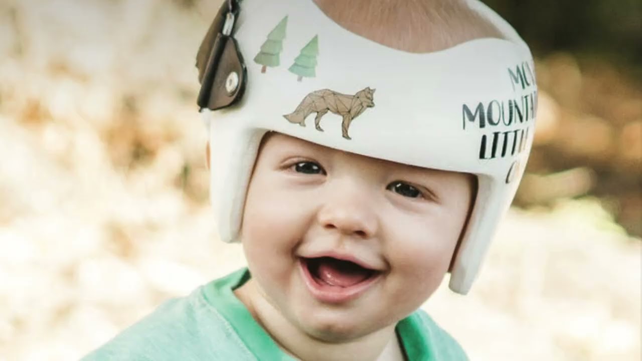
What causes Craniosynostosis?
In most children, the exact cause is unknown.
About 20% of cases occur as part of a genetic condition or craniofacial syndrome, such as Apert syndrome or Crouzon syndrome. Children with these syndromes are more likely to have more than one skull suture close too early and may have other differences affecting the face, hands, feet, eyes, ears or airway.
Because craniosynostosis can affect multiple parts of the body, children often benefit from care provided by a team of many pediatric specialists who work together to create a personalized treatment plan.
Researchers continue to study what causes craniosynostosis. While some genetic changes have been linked to the condition, the exact reason it develops in many children is still not fully understood.
Would you like to schedule an appointment with Neurosurgery?
What are the signs and symptoms?
The most noticeable sign is an unusual head shape that care teams or parents see at birth or become more noticeable as a baby grows. The exact shape depends on which skull suture closes too early.
Parents or pediatricians may notice:
- A long, narrow or uneven head shape
- A forehead that looks pointed, flat or more prominent on one side
- One side of the face or eye socket appearing different from the other
- A raised ridge along a skull suture
- A soft spot that feels unusual or seems to close early
- Little or no change in head size as the baby grows
Many babies otherwise appear healthy and may not have pain or other obvious symptoms. In more complex cases, restricted skull growth can increase pressure around the brain. Signs may include vomiting, unusual sleepiness, irritability, vision problems or developmental concerns.
An unusual head shape can mean many different things. Positional plagiocephaly, a common and usually harmless flattening of the head, can look similar. A craniofacial specialist can determine the cause and whether treatment is needed.
How is Craniosynostosis treated?
Most children with craniosynostosis will need surgery to allow the skull to grow normally as the brain develops and improve the shape of the head. However, very mild cases may not require surgery.
When is surgery performed?
When surgery is recommended, it is often performed during the first year of life, when an infant's skull is growing rapidly and the bones are more flexible. Early diagnosis and evaluation help ensure your child has the greatest range of treatment options.
Which type of surgery is right for my child?
There are several successful options for surgeries. The right procedure depends on your child's age, the type of craniosynostosis and their overall medical condition.
Because no single surgical approach has been shown to provide better cosmetic or functional outcomes than another, our philosophy is to recommend the least invasive procedure that is appropriate for your child's condition.
Surgical options
- Endoscopic surgery: Babies younger than 6 months may be candidates for this minimally invasive approach.
- Open cranial reconstruction: Older infants and children more commonly benefit from this procedure, which is performed by our pediatric neurosurgery and plastic surgery craniofacial team.
Your child's surgeon will carefully evaluate their individual needs and recommend the approach that's right for them.
Surgery to correct craniosynostosis can be performed in several ways, depending on the age of the patient and the type of deformity. No single method has been shown to produce superior cosmetic or functional outcomes, and so our philosophy is to always employ the least invasive surgical technique appropriate for that child’s deformity. Children under 6 months of age often have a minimally invasive, endoscopic surgery. Older children will more commonly benefit from an open cranial reconstruction, performed by our joint plastic surgery and neurosurgery craniofacial team. But age alone is not the only factor in determining who is eligible for either procedure: Your craniofacial surgeon will consider your child’s entire medical condition before making their recommendation.
Long Term Care for Craniosynostosis
Late complications after surgical repair of craniosynostosis are rare, but your child will require continued monitoring for several years after surgery to look for late impacts to vision, secondary fusions of the skull, persistent skull defects, and trends in their neurodevelopment. Patients whose craniosynostosis was part of an overarching syndrome may require additional screening or treatment for other abnormalities of the face and ears as well.
Our multidisciplinary Craniofacial Clinic allows families to be seen by expert pediatric and surgical specialists from all craniofacial disciplines in a single location, providing comprehensive and convenient pre-and post-surgical care from birth to young adulthood.